Osteoid osteoma (OO) is the most common benign osteogenic bone neoplasm and occurs predominantly in young, male patients [1]. The preferential site of this neoplasm is the cortex of long bones, especially the femur and the tibia [2]. OO is typically characterized by nocturnal pain and response to non-steroidal anti-inflammatory drugs (NSAIDs). Radiological characteristics are a relatively radiolucent small nidus within an area of extensive reactive sclerosis [3]. It is important to know that in some cases, this neoplasm cannot be appropriately individualized with conventional imaging methods such as radiography resulting in diagnosis difficulties [4, 5].
Its localization in the toe is rare and can be difficult to diagnose especially if associated with swelling and erythema. We report the case of a woman with OO of the distal phalanx of the hallux, initially diagnosed as metatarsalgia and inappropriately treated.

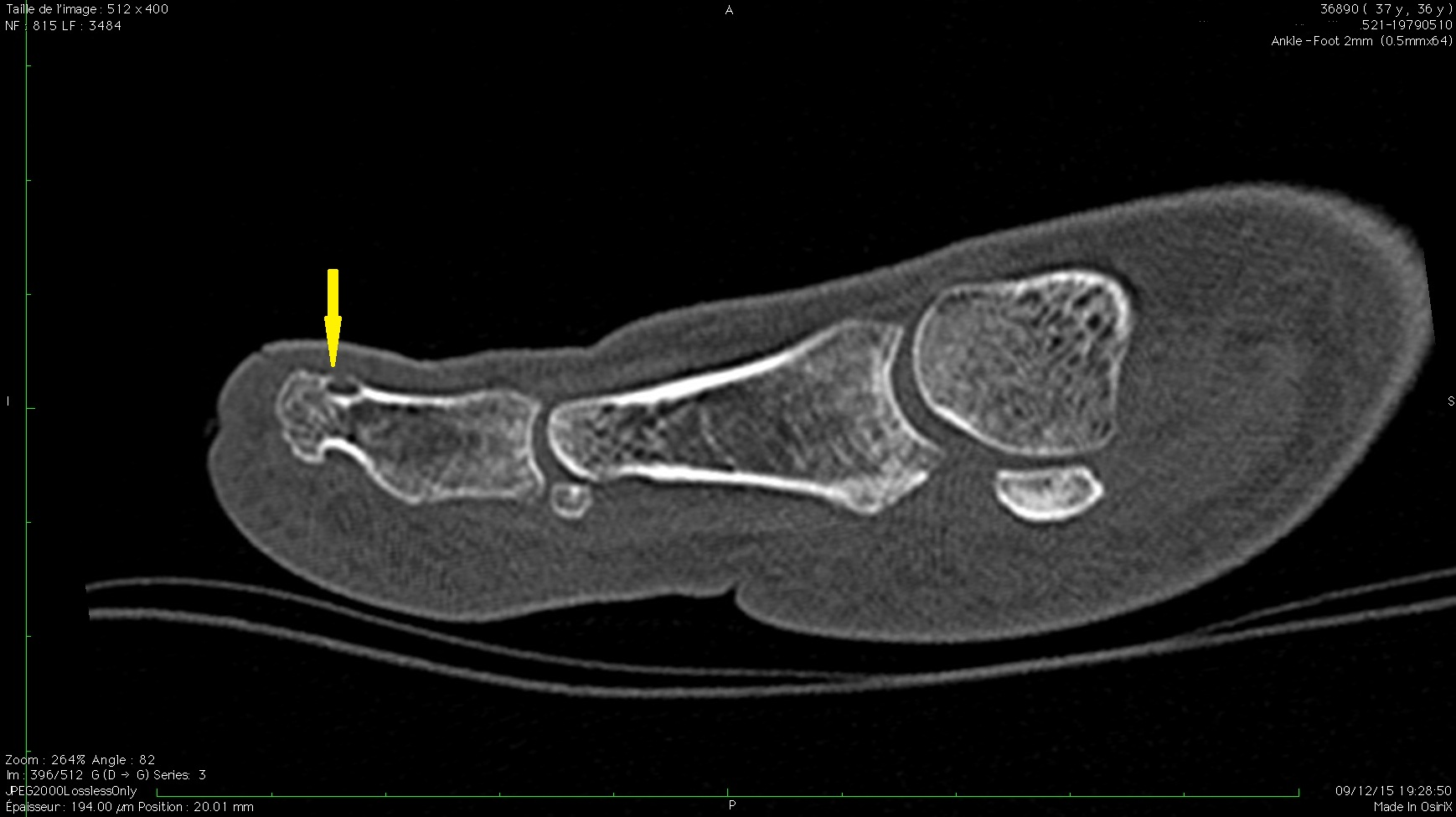
A 37-year-old dentist woman was complaining of a progressive moderate pain of the right great toe with no trauma history. As the pain was aggravated with standing posture, weight bearing and press on the pedal of the dentist chair while working, her therapist initially retained the diagnosis of metatarsalgia and she received only painkillers. Six months later, she presented to the outpatient of Physical Medicine and Rehabilitation with the same complaint reporting that pain worsened, especially at night. Physical examination revealed a moderate swelling and tenderness on palpation of the distal phalanx of the hallux. There was no hyperemia or temperature increase. The hallux exhibited no skin or nail abnormalities. The C-reactive protein level, the white blood cell and the erythrocyte sedimentation rate were within normal limits. X ray of the foot showed a faint radiolucent area in the tip of the distal phalanx of the hallux. The diagnostic of osteoid osteoma was suspected, NSAIDs were prescribed and pain was alleviated. CT scan revealed the presence of a small lytic lesion with peripheral cortical thickening (LODWICK1A) measuring 3mm in the dorsal side of the second phalanx of the right hallux (Figure 1). MRI examination showed hyperintense STIR and T2 signal (Figure2), T1 hypointense, with contrast uptake after injection. The patient refused surgical or radiofrequency ablation. Her symptoms resolved under NSAIDs with no signs of recurrence at two years follow up.
OO of the foot is a rare condition. A systematic review published in 2015 revealed that the talus was the most commonly affected bone in the foot, whereas phalanx of the toe localization accounts for 2% of all cases [6]. To the best of our knowledge, only few cases reporting OO of the distal phalanx of the toes are published in the current literature [7-10]. The unusual location in the foot of OO can be confused with chronic arthritis, twisted ankle, os trigonum syndrome or osteomyelitis, resulting in a delay of the diagnosis and the operative management.
Phalangeal lesions are typically small and are associated with local tenderness. Soft tissue swelling and aching pain with nail hypertrophy may occur when the lesion affects the distal phalanx [10].
The radiologic presentation of OO of the foot is characterized by a mild to absent osteosclerosis which may be located at a distance from the nidus. [11] The juxta-articular situation of the neoplasm can make the diagnosis harder especially if the sclerosis is absent [3]. When the diagnosis is suspected, it is imperative to continue the investigation with bone scintigraphy, CT scan or MRI [12].
CT scan is considered to be superior to MRI in the diagnosis and evaluation of OO. It can also be used to guide percutaneous removal of the nidus [12, 13]. When the nidus cannot be appropriately identified, MRI is particularly useful showing the bone marrow edema signal [12]. Typically an early and intense Gadolinium enhancement, is found in relation to the nidus hyper vascularization [14].
OO treatment is based on the removal of the entire nidus. As our patient refused surgical treatment or radiofrequency ablation, she received NSAIDs resulting in complete remission of her symptoms with no recurrence at two years follow up.
Similar cases of medical management with NSAIDs leading to resolution of the disease were previously reported in the literature. Nevertheless, the long-term effects and recurrence rates have not been clearly documented [15]. Surgical procedures include open surgical excision and percutaneous techniques consisting of ethanol injection [16], radiofrequency ablation [17], and laser photocoagulation [18]. As for conservative treatment with NSAIDs, the main drawback of the percutaneous procedures is the impossibility of histopathologic confirmation.
Localization of OO on the distal phalanx of the hallux remains a rare medical condition, illustrating the difficulties associated with the early diagnosis of the lesion.
This case highlights the fact that OO should be suspected in a patient with a chronic foot pain that changes to become nocturnal and disappears with NSAIDs administration. A combination of medical history, a detailed assessment of both clinical and radiological features should lead to the correct diagnosis. In our case, histopathological confirmation was impossible as the patient refused to undergo surgery. NSAIDs administration resulted in complete remission of her symptoms with no recurrence at two years follow up. Otherwise, failure or intolerance of the medical management would probably have led to open surgical resection or percutaneous radiofrequency ablation.
- Onoue K, Kudawara I. Osteoid osteoma with cartilage formation of the distal phalanx in the toe. Orthopedics. 2007;30:670-1 pubmed
- Sproule J, Khan F, Fogarty E. Osteoid osteoma: painful enlargement of the second toe. Arch Orthop Trauma Surg. 2004;124:354-6 pubmed
- Davies M, Cassar Pullicino V, Davies A, McCall I, Tyrrell P. The diagnostic accuracy of MR imaging in osteoid osteoma. Skeletal Radiol. 2002;31:559-69 pubmed
- Ilaslan H, Sundaram M. Advances in musculoskeletal tumor imaging. Orthop Clin North Am. 2006;37:375-91, vii pubmed
- Nakamura SA, Lorenzato MM, Engel EE, Yamashita MEAS, Nogueira-Barbosa MH. Incidental enchondromas at knee magnetic resonance imaging: intraobserver and interobserver agreement and prevalence of imaging findings. Radiol Bras. 2013;46(3):129-33.
- Kneisl J, Simon M. Medical management compared with operative treatment for osteoid-osteoma. J Bone Joint Surg Am. 1992;74:179-85 pubmed
- Akhlaghpoor S, Tomasian A, Arjmand Shabestari A, Ebrahimi M, Alinaghizadeh M. Percutaneous osteoid osteoma treatment with combination of radiofrequency and alcohol ablation. Clin Radiol. 2007;62:268-73 pubmed
- Cioni R, Armillotta N, Bargellini I, Zampa V, Cappelli C, Vagli P, et al. CT-guided radiofrequency ablation of osteoid osteoma: long-term results. Eur Radiol. 2004;14:1203-8 pubmed
- Witt J, Hall Craggs M, Ripley P, Cobb J, Bown S. Interstitial laser photocoagulation for the treatment of osteoid osteoma. J Bone Joint Surg Br. 2000;82:1125-8 pubmed